AI-Based CT Can Accurately Screen Patients for TMVR
The fully automated method could increase clinical workflow efficiency and potentially aid trial enrollment, says Martin Beyer.

LISBON, Portugal—Using artificial intelligence (AI)-based CT to screen patients for transcatheter mitral valve replacement (TMVR) is as reliable as screening that uses the traditional core lab approach, according to new data.
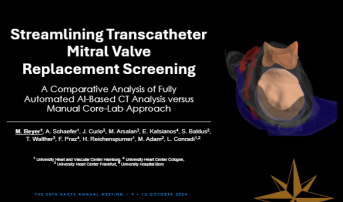
Martin Beyer, MD (University Heart and Vascular Center, Hamburg, Germany), who presented the findings at the European Association for Cardio-Thoracic Surgery 2024 meeting, told TCTMD that AI-based CT screening in the clinic can save time and resources.
Patients evaluated for TMVR tend to have a high burden of disease with challenging traits like mitral annular calcification and prior valvular intervention, which can make preprocedural measurements needed for clearance tricky, he explained. In fact, screen failure rates have remained “high,” between 60% and 89% in studies conducted over the last 5 years.
“Extensive CT analysis and high screen failure rates hinder broader applicability for TMVR, resulting in time- and resource-intensive screening,” Beyer said during his presentation. From a research perspective, he added, the AI-based CT analysis could also aid in increasing clinical trial enrollment.
Software-Informed vs Manual Analysis
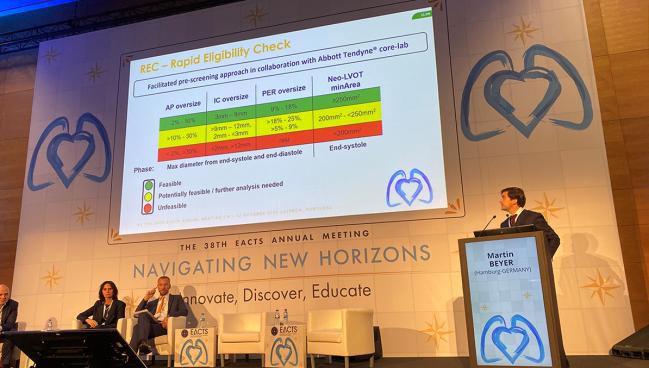
The study included 181 patients screened for TMVR with Tendyne (Abbott) at one of four high-volume centers in Germany and Switzerland. Cardiac computed tomography angiography (CCTA) scans from patients with severe mitral regurgitation were reviewed conventionally with a manual core lab CT analysis as well as with a fully automated AI-based method. With AI software (LARALAB) that uses a novel tool called rapid eligibility check, TMVR is categorized as feasible, potentially feasible/further analysis needed, and unfeasible in a matter of about 15-20 minutes based on several measurements.
Beyer and colleagues found “really good agreement” for all parameters analyzed via AI or the conventional approach, he said. These included mitral annular perimeter, intercommisural distance, septolateral diameter, and minimal end-systolic left ventricular outflow tract area.
The CT-AI performed with a sensitivity of 91.6% and a specificity of 94.2% compared with the manual approach, with 8% false negatives and 6% false positives.
“All in all, really, really promising, really decent results,” Beyer said. He noted that the AI-assessment is limited to gauge anatomical eligibility and that patients may still be ineligible based on other clinical factors. Additionally, it has so far only been validated against the core-lab screening method and not yet for its performance in real-world clinical settings.
Arnaud Van Linden, MD, PhD (Saarland University Medical Center, Homburg, Germany), who served as the session moderator, said he uses the same software for type A aortic dissection screening so might be biased. That said, “I'm really sure that this will be the future in CT imaging or even echo imaging or whatever to help interpret the images and also screen patient,” he predicted.
To TCTMD, Van Linden said the results he’s seen with this software are “really amazing,” especially since the AI-based system can be accessed at any time and from anywhere. The tool is “more or less” ready to be used in clinical practice once it receives regulatory signoff, he said. Reimbursement is an issue still because the technology is cloud-based, “but from a technical point of view, we could start tomorrow to implement it.”
Given growing concerns with data security, Van Linden also acknowledged the need for good IT support, though he noted this challenge is not unique to this particular AI product.

Yael L. Maxwell is Senior Medical Journalist for TCTMD and Section Editor of TCTMD's Fellows Forum. She served as the inaugural…
Read Full BioSources
Beyer M. Streamlining transcatheter mitral valve replacement screening: a comparative analysis of fully automated AI-based CT analysis versus manual core-lab approach. Presented at: EACTS 2024. October 10, 2024. Lisbon, Portugal.
Disclosures
- Beyer reports receiving travel support from Abbott and research grant support from the German Heart Foundation.
Related News


Comments