Probe Beyond Ejection Fraction to Understand and Manage HFpEF
Fibrosis and arterial stiffness, for example, differed in patients with EF under or over 60%, offering clues for care.

Among patients with heart failure with preserved ejection fraction (HFpEF), lower versus higher ranges of EF signal fundamentally different morphologies and hemodynamic responses, which may have implications for pharmacologic management, new data confirm.
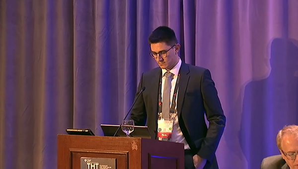
“[W]e propose that therapies addressing stress volume and ventricular fibrosis might be more favorable in low LVEF [HFpEF] patients, while therapies addressing arterial stiffness might be more effective in HFpEF with high LVEF,” said Sebastian Rosch, MD (Heart Center Leipzig, Germany), during a late-breaking clinical science presentation at the Technology and Heart Failure Therapeutics (THT) 2022 meeting in New York.
Rosch noted that in the EMPEROR-preserved trial, which showed benefit of the sodium-glucose cotransporter 2 (SGLT2) inhibitor empagliflozin (Jardiance; Boehringer Ingelheim/Eli Lilly) in HFpEF patients with or without diabetes, the drug did not work as well in those in the highest EF range (60% or higher), an attenuation that had been observed in other HFpEF trials as well. At the time EMPEROR-preserved was presented, commentators floated the idea that heart failure categories might need to be renamed. And while “midrange” EF has been used in the past for ejection fractions between 40 and 49%, the recent European Society of Cardiology guidelines proposed that this category be renamed “mildly reduced.”
A deep dive into HF phenotypes would be one way to parse the differences. For the current study, Rosch and colleagues set out to investigate whether stratification of HFpEF patients by LVEF status identifies differential morphological and functional subphenotypes.
Using three different phenotyping measures, Rosch and colleagues found that patients with an EF in the range of 50% to 60% were technically more like HF with reduced ejection fraction (HFrEF) patients in terms of ventricle size, extent of fibrosis, and impaired ventriculoarterial coupling. HFpEF patients on the higher end of the EF range, on the other hand, had high end-systolic elastance on handgrip exercise and a marked preload vulnerability.
Marked Differences Emerge
For their study, Rosch and colleagues enrolled 56 HFpEF patients. Of these, 21 patients had an EF in the range of 50% to 60%, while 35 had an ejection fraction greater than 60%. Phenotyping included histology on myocardial biopsy, echocardiography and cardiac MRI, and invasive hemodynamic characterization via pressure volume loop analysis.
Compared with those with lower ranges, the group of patients in the EF 60% or higher cohort were slightly older and more likely to be female, while those in the low-EF group had higher rates of diabetes and past or current smoking. Baseline N-terminal pro-brain natriuretic peptide (NT-proBNP) levels, however, did not differ between the two groups. In terms of clinical presentation, the groups also had similar blood pressure measurements at admission, as well as similar representation of patients in NYHA classes II, III, and IV. Echocardiographic estimates of ventricular hypertrophy and filling pressures also showed no significant differences between groups.
When myocardial fibrosis was quantified on histology in a subgroup of 28 patients, the lower-range EF group had a greater mean extent of fibrosis than the high-EF group (15.2% vs 9%). On cardiac MRI, end-diastolic volumes were higher in the low-EF group (78 mL/m2 vs 68 mL/m2 in the high-EF group), as were end-systolic volumes (38 mL/m2 vs 25 mL/m2). In this subgroup, the mean EF was 54% in the low-EF group and 64% in the high-EF group. While no differences were seen in left ventricular stroke volume, myocardial mass was higher in the low-EF cohort (63 g/m2 vs 54 g/m2), but relative wall thickness was higher in the high-EF cohort (0.80 vs 0.73).
In the pressure volume loop analysis, Rosch and colleagues found higher end-systolic and end-diastolic pressures in the high-EF group, as well as higher systolic and diastolic stiffness, compared with the low-EF group. During handgrip exercises, the low-EF cohort showed left ventricular pressure shifts typical of patients with HFrEF. In contrast, the high-EF cohort had a pronounced increase in effective arterial elastance, suggestive of higher arterial resistance and an implication of higher afterload sensitivity.
“Patients with high EF do not only appear to be afterload sensitive, but also preload sensitivity,” Rosch said. “During preload reduction during transient inferior caval vein occlusion, we see a dramatic drop of left ventricular pressures and volumes, and consequently we see a relevant drop in stroke volume, demonstrating preload susceptibility in high EF patients.”
Panelist Clyde Yancy, MD (Northwestern University Feinberg School of Medicine, Chicago, IL), commenting on what he called a “very elegant physiologic study,” noted that further information might be gleaned by replicating the analysis and switching out EF for a cardiac MRI-derived strain measurement.
Another panelist, Anu Lala, MD (Icahn School of Medicine at Mount Sinai, New York, NY), noted that emerging data point toward exercise intolerance being a function of aortic stiffness in HFpEF patients, particularly in women. Similarly, co-moderator JoAnn Lindenfeld, MD (Vanderbilt University Medical Center, Nashville, TN), stressed the importance of gender analyses in phenotype research.
Rosch agreed, noting that much more research is needed to better understand how pharmacologic therapies can best address arterial stiffness.
Commenting on the study for TCTMD, Alexander Hajduczok, MD (Thomas Jefferson Hospital, Philadelphia, PA), said while it’s too soon to say that high- and low-range HFpEF patients should be treated differently in terms of pharmacotherapy, the data are hypothesis-generating, both from a hemodynamic standpoint and from a big-picture perspective of moving toward understanding why some patients benefit more from a given therapy than others.
“I think that you'd probably need a larger cohort to really nail down more specific hemodynamic changes, but this is a good first step in trying to do that,” he said. Like Yancy, Hajduczok said EF is just one metric, which may or not be the best one for getting to the bottom of this issue.
“The field in general is still working out the kinks of how to correctly identify or phenotype within HFpEF,” he said. “Some people may have a completely normal EF, some have a slightly reduced EF, and others may have differences in inflammation, or systemic disease, or potentially amyloid. You also have to consider how decompensated people are, and how high their filling pressures are, etc. But, showing marked differences among these two subgroups [is] a step in the right direction.”
L.A. McKeown is a Senior Medical Journalist for TCTMD, the Section Editor of CV Team Forum, and Senior Medical…
Read Full BioSources
Rosch S. Hemodynamic Profiles across the range of left ventricular ejection fraction in heart failure with preserved ejection fraction—potential therapeutic implications. Presented at: THT 2022. New York, NY. February 2, 2022.
Disclosures
- Rosch reports no relevant conflicts of interest.


Comments