New Expert Advice on Intracoronary Imaging Urges Shift to Preprocedural Planning
Cost and time are two oft-cited reasons for not imaging the coronaries. But lack of expertise and education shoulder some blame.

And while add-on procedural time and economic considerations are often cited as the chief barriers to performing IVUS or optical coherence tomography (OCT) imaging, experts say the biggest barrier is likely the lack of physician expertise in interpreting the imaging results.
“Cost, both time and financial, are two of the greatest restrictions” to wider adoption of intracoronary imaging, lead author Thomas Johnson, MD (Bristol Heart Institute, England), told TCTMD. “But there’s also an unwillingness, interestingly, on the part of the interventionalists to expose themselves as not understanding the technology and that’s a real, real issue.”
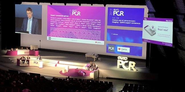
The new statement follows a “Part 1” document, presented and published at EuroPCR last year, addressing the use of intracoronary imaging to enhance patient selection and stent optimization. Part 2, presented by Johnson yesterday at EuroPCR, was simultaneously published online in the European Heart Journal and endorsed by the Chinese Society of Cardiology, the Hong Kong Society of Transcatheter Endocardiovascular Therapeutics (HKSTENT), and the Cardiac Society of Australia and New Zealand.
But there’s also an unwillingness, interestingly, on the part of the interventionalists to expose themselves as not understanding the technology and that’s a real, real issue. Thomas Johnson
Johnson said it was “gratifying” to see, following the publication of last year’s document, that the latest European guidelines for myocardial revascularization upgraded the recommendations for OCT to class IIa for the optimization of stent placement, matching the recommendation for IVUS in this context. IVUS can also be considered for the assessment of lesion severity for unprotected left main lesions (class IIa), and both IVUS and OCT “should be considered” for the detection of stent-related mechanical problems leading to restenosis, again a class IIa recommendation in the latest guidelines.
More evidence in support of intracoronary imaging has also emerged in the interim. This was ULTIMATE, released at TCT 2018, showing a 2.5% absolute reduction in the risk of target vessel failure among patients whose DES implantation was IVUS-guided as compared with patients whose procedures were planned and executed based on angiography alone.
Recommendations, Sparse Evidence
But putting together the recommendations for Part 2 has been “more troublesome,” Johnson acknowledged during a EuroPCR press conference, due the lack of published data in this area. Despite the need for more randomized trials in this space, the writing group advocates a “shift in focus” such that intracoronary imaging is considered not merely as a procedural tool during stent implantation, but also as a means to better understand lesion morphology during the planning stages—specifically, the composition of the atherosclerotic plaque, detection of culprit lesions, and markers of vulnerability.
To synthesize the information in their document, Johnson and colleagues provide boxed summaries for three key themes:
- Indications and clinical value of intracoronary imaging in ACS
- Role of imaging in vulnerable plaque and risk stratification
- Consensus recommendation on the role of imaging to assess lesion significance
Also provided is a proposed algorithm for the use of intracoronary imaging in a patient with suspected ACS.
“We really see imaging as central to . . . understanding the etiology of acute coronary events, particularly in the nonobstructive or near-normal coronary angiogram,” Johnson told reporters. “Beyond that, in terms of vulnerable plaque detection and risk stratification, there appears to be evidence that a multimodality approach to understand the underlying plaque type is probably relevant in terms of intracoronary imaging.”
Taken together, he said, the two intracoronary imaging consensus documents stem from an acknowledgement that operators are seeing increasingly complex patients and lesions.
“Inherently, and particularly emphasized in this second document, we understand that invasive angiography is flawed in terms of its assessment and we should be shifting towards a more precision-based medical strategy for our patients,” Johnson continued. “Intracoronary imaging allows lesion characterization, as expanded upon in our document, and stent selection and stent optimization [covered in Part 1]. This will facilitate for us a tailored approach to intervention from the preintervention strategy to subsequent stent deployment.”
Use Begets Use
Speaking with TCTMD, Johnson stressed that uptake of intracoronary imaging varies substantially by region, ranging from more than 80% in Japan to just 5% in the United Kingdom. An EAPCI/Cardiovascular Interventions and Therapeutics survey, he noted, suggests that operators with more years of experience are also more likely to be the ones using more intracoronary imaging.
“So anecdotally, that emphasizes that the more you use imaging, the more you realize that you can’t rely upon your interpretation of the angiogram,” he said. “It’s self-fulfilling, but the more we do, the more actually we need to see and rely upon the information we gain from the intracoronary imaging modalities.”
Johnson agreed that financial costs and time pressures play a significant role in an operator’s decision to proceed with IVUS or OCT, but he reiterated the need for operators to read the consensus documents and appreciate what they’re missing out on. “The challenge is two-fold,” he said. “We have an absolute duty to better understand the complexity of disease, because without that we’re never going to achieve optimal results, but we also have a massive element of education to work on in terms of fostering confidence in the interventional community to undertake the imaging. And that’s probably the biggest barrier.”
He added that he thinks there has been a groundswell of interest in intracoronary imaging in the past year, in part because of the guideline upgrades, in part because of ULTIMATE, but also because of the advent of new technologies like lithotripsy balloons, which require understanding of the underlying plaque composition. “Unless you image and understand what you’re dealing with, how can you choose a lithotripsy balloon over a noncompliant balloon over a rotablator? We have to be tailoring our treatment, and we’re only going to achieve that if we know what we’re dealing with, so we have to look.”
Shelley Wood was the Editor-in-Chief of TCTMD and the Editorial Director at the Cardiovascular Research Foundation (CRF) from October 2015…
Read Full BioSources
Johnson T, Räber L, di Mario C, et al. Clinical use of intracoronary imaging. Part 2: acute coronary syndromes, ambiguous coronary angiography findings, and guiding interventional decision-making: an expert consensus document of the European Association of Percutaneous Cardiovascular Interventions. Eur Heart J. 2019;Epub ahead of print.
Disclosures
- Johnson reports participating in Abbott’s speaker’s bureau and receiving grant/research support from AstraZeneca and honoraria/consulting fees from Abbott, Bayer AG, Biosensors, Boston Scientific, Medtronic, and Terumo.


Comments