Preprocedural AI-QCA Matches OCT-Guided PCI for Noncomplex Lesions
Several experts argue that AI-QCA is not a replacement for OCT, but could be valuable in low-resource settings.

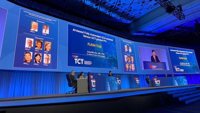
WASHINGTON, DC—Using an artificial intelligence (AI)-based quantitative coronary angiography (QCA) to help plan PCI may be as good as optical coherence tomography (OCT) guidance in achieving good minimal stent area (MSA) for noncomplex lesions, according to new randomized data from Korea.
Experts continue to advocate for increased use of intravascular imaging to aid PCI in real time with plenty of data to back up this practice, especially in complex lesions, but many cath labs have yet to embrace the technology, usually relying on angiography alone to plan procedures. QCA has been used even less often than intravascular imaging in contemporary practice, but previous results from GUIDE-DES suggested the former could be an alternative to the latter in certain situations, especially given its lower cost.
“AI-QCA-assisted PCI achieved a noninferior minimal stent area compared to the OCT-guided PCI in simpler procedural safety and in 6-month outcomes,” said Jung-Min Ahn, MD (Asan Medical Center, University of Ulsan College of Medicine, Seoul, Republic of Korea), who presented the FLASH findings today at TCT 2024. “AI-QCA offers a very valuable solution, especially in a resource-limited setting or in less complex cases where intravascular imaging may have a limited benefit.”
He advocated for larger trials tracking long-term clinical outcomes to better “establish AI-QCA’s role in routine interventions.”
The study was simultaneously published in JACC: Cardiovascular Interventions with first author Yongcheol Kim, MD (Yonsei University College of Medicine and Cardiovascular Center, Yongin Severance Hospital, Republic of Korea).
“AI-QCA could have a really important impact on places where they're not using [OCT]; it's probably better than just using an eyeball,” said Roxana Mehran, MD (Icahn School of Medicine at Mount Sinai, New York, NY), during a press conference, stressing that it cannot be a replacement for intravascular imaging. “We finally have data that proves that intravascular imaging can have an important impact on outcome measures, [and that] can't be understated.”
During the session, panelist Mamas Mamas, BMBCh, DPhil (Keele University/Royal Stoke University Hospital, Stoke-on-Trent, England), TCTMD’s Senior Clinical Editor, agreed that OCT should not be swapped for AI-QCA. However, “in low-resource settings, it's useful,” he said.
Speaking with TCTMD, Javier Escaned, MD, PhD (Hospital Clínico San Carlos, Madrid, Spain), said he feels similarly. In his experience with the system used in the study, it “simplifies enormously the task of obtaining very accurate measurements,” said Escaned. As such, the “era of eyeballing PCI is coming to an end or should be coming to an end.”
Noninferiority Criteria Met
For the study, Ahn and colleagues randomized 400 patients (mean age 65.1 years; 81.8% men) from 13 centers in Korea to undergo fully automated AI-QCA prior to PCI (MPXA-2000 system; Medipixel) or OCT-guided PCI between October 2022 and February 2024. Patients were excluded if they had left main, chronic total occlusion, graft vascular, or bifurcation lesions as well as if the OCT catheter could not cross the lesion.
Patients in the AI-QCA arm underwent unblinded post-PCI OCT evaluation, and while discouraged, further procedures were allowed to correct suboptimal stent results. Mean stent diameter was slightly larger in the AI-QCA group (3.25 vs 3.0 mm; P = 0.025) as was maximal noncompliance balloon size (3.7 vs 3.6 mm; P = 0.040).
There was no difference in the primary endpoint of post-PCI MSA between the AI-QCA and OCT-guided PCI groups (6.3 vs 6.2 mm2) which met the criteria for noninferiority with a margin of 0.8 mm2 (P < 0.001) but not superiority (P = 0.48). The results were consistent in sensitivity analyses looking at segment type and side branch.
There were no differences between the study groups in terms of the OCT-defined endpoints of overall stent expansion, stent underexpansion, dissection, and untreated reference segment disease, but there was more malapposition in the AI-QCA arm (13.6% vs 5.6%; P = 0.007). Angiographic endpoints and procedural complications were also all similar between AI-QCA and OCT. However, 16.5% of patients in the AI-QCA arm received either additional ballooning for malapposition (n = 5) or underexpansion (n = 26) or additional stenting for dissection (n = 2) after the primary endpoint was evaluated.
Clinical outcomes at 6 months were comparable. There was a single noncardiac death in the AI-QCA arm as well as one target vessel revascularization in the OCT cohort.
Ahn acknowledged the study’s limitation of using MSA as a surrogate marker rather than evaluating a clinical endpoint. Additionally, he said, “low complication rates at 6 months limited definitive conclusions about procedural safety.”
The results suggest that “AI-QCA technology can be effectively integrated into routine PCI, bridging conventional angiography and imaging-guided PCI,” Ahn concluded.
Discussing the study during the session, Akiko Maehara, MD (Columbia University and Cardiovascular Research Foundation, New York, NY), said FLASH “may change” current practice in that AI-QCA is “better than visual estimation of vessel size or stenosis evaluation.”
A major strength of the study is that MSA is “the most robust single parameter to be associated with long-term clinical outcome.” She acknowledged prior criticism of the noninferiority margin used in the study being potentially too wide but called it “appropriate.”
Going forward, Maehara noted, the proper lesions and operators for AI-QCA remain unclear. “Operators in the current trial have imaging trained eyes to interpret angiography, thus applicability for nonimaging operators are unknown,” she added.
‘The First Step’
The advantages of AI-QCA over manual QCA are that it is “easier to obtain and faster,” Escaned said, adding that the results of FLASH might “lead to a rebirth” of this technology, which has “disappeared out of the mental map of interventional cardiology.”
However, it remains to be seen how AI-QCA performs in more complex lesions that have shown to especially benefit from intravascular imaging, he added. In “no way” could it ever replace OCT, Escaned continued, given that it gives no information regarding vessel wall characteristics that could guide ideal stent placement.
If AI could eventually provide functional information from the angiogram, “this could be a revolutionary way of performing PCI,” Escaned observed.
During the press conference, Joost Daemen, MD, PhD (Erasmus University Medical Center, Rotterdam, the Netherlands), said that he welcomes the addition of AI-based image interpretation into clinical practice. “That is where the future lies in helping us more easily interpret the severity, plaque recognition, and so on,” Daemen observed. “It's extremely interesting that solutions like this are becoming available, are becoming more easily applicable and, even online. That's a very good thing.”
However, he did take issue with a comparison of a pre-PCI-only assessment tool with image-guided PCI. “That bypasses the whole concept of image-guided PCI in my perspective,” Daemen argued, adding that research over the past decade has shown that the value of intracoronary imaging comes from not only intraprocedural assessment but also post PCI feedback. “I cannot envisage how just a pre-PCI assessment, the OCT or this, can be just as good as image-guided PCI. So that makes me feel a bit uncomfortable.”
Ahn agreed, explaining that this is why high-risk patients such as those with left main disease were not included in the study. “We can’t apply it in the whole [spectrum of] lesion subsets including complex lesions,” he said.
The role of AI across the field of interventional cardiology is to “democratize procedures,” said Susheel Kodali, MD (NewYork-Presbyterian/Columbia University Irving Medical Center, New York, NY), during the press conference. “AI is a cool term, but it has to improve efficiency, safety, or access and availability. And I think technologies like this can, we just have to validate them, and I struggle with how to validate some of these measures.”
Ahn called the findings “the first step.” As the technology improves, “we can apply it to other people,” he added.
In an accompanying editorial, Mohamad Alkhouli, MD, and Shih-Sheng Chang, MD, PhD (both Mayo Clinic, Rochester, MN), write that FLASH “could serve as a foundation for a future, larger trial, powered for clinical outcomes.”
Ultimately, they say, “the extent to which AI can democratize optimal care will largely depend on how intuitive and user friendly the technology is for routine use.”
“Although much work remains, we are steadily progressing toward a future when comprehensive AI solutions will play a central role in diagnosing and treating CAD in the catheterization laboratory,” the editorialists conclude.
Yael L. Maxwell is Senior Medical Journalist for TCTMD and Section Editor of TCTMD's Fellows Forum. She served as the inaugural…
Read Full BioSources
Kim Y, Yoon H-J, Suh J, et al. Artificial intelligence-based fully automated quantitative coronary angiography versus optical coherence tomography guided PCI (FLASH trial). J Am Coll Cardiol Interv. 2024;Epub ahead of print.
Alkhouli M, Chang S-S. AI-assisted PCI: progress, hurdles, and future pathways? J Am Coll Cardiol Interv. 2024;Epub ahead of print.
Disclosures
- This project was supported by the Korea Medical Device Development Fund grant funded by the Korea government and Medipixel.
- Ahn reports receiving consulting fees from Medipixel.
- Kim, Escaned, Alkhouli, and Chang report no relevant conflicts of interest.


Comments