Going Contrast Free in TAVI Is Safe for CKD Patients
Promising data for noncontrast CCTA are starting to build, with the latest study showing its role in balloon-expandable valves.


With the right imaging strategy—one that avoids contrast media—patients with advanced chronic kidney disease (CKD) can safely undergo TAVI without putting undue strain on their renal function, a new study confirms.
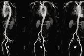
In a paper published recently in JSCAI, David Elison, MD, and colleagues at the University of Washington in Seattle describe their approach to contrast-free TAVI: an algorithm that can gauge balloon-expandable valve size based on sinus and sinotubular junction diameters obtained from noncontrast coronary CT angiography (CCTA), adapted for patient sex.
“Iodinated contrast exposure may worsen kidney function in a dose-dependent fashion, and may result in a need for renal replacement therapy. Therefore, a method to eliminate, or greatly lessen, contrast exposure during TAVR is highly desirable,” the researchers note, pointing out that poor or worsening renal function, both before and after the valve intervention, are linked to higher mortality risk.
Senior author James M. McCabe, MD (University of Washington), told TCTMD that these scenarios where going no contrast might be helpful, while in the minority, are common “enough that it became important for us to try and nail down a methodology.”
With growing expertise, the segment of patients considered candidates also grows, he added. “In the beginning you might think that this sort of strategy is a bit high risk and feels a little outside your comfort zone, and so you might just pick patients who are really just at the precipice of needing dialysis. But as the technique becomes more comfortable for an operator, that threshold . . . starts to go down.”
Alternatives to noncontrast CCTA for TAVI do exist, McCabe noted. One option is transesophageal echocardiography (TEE), which “is often referred to as the gold standard but hasn’t actually been validated in contemporary times all that well,” he said. As TAVI is expanded to more and more centers, each imager may gain less experience with that technique, he added. The other option is cardiac MRI, “which I think can be instituted for elective patients but is awfully hard to do, if not impossible to do, in patients who are in shock or in some extremis,” a population that tends to have higher prevalence of comorbidities like CKD.
For their center, “trying to come up with a streamlined process was important,” said McCabe, who initially started doing the CT-based strategy back in 2019. Over time, he and his colleagues sought to develop an algorithm that was “teachable and transparent.”
Whatever approach is used to cut back on contrast, be that CTA, MRI, or TEE, the main point is it’s “feasible and that we need to continue to emphasize the importance of renal protection for patients at risk,” McCabe stressed. “Ultimately, institutions can choose between the multiple plausible methodologies that have been advanced in a way that best matches the strengths of their program.”
Pedro Nicz, MD (Heart Institute - InCor/HCFMUSP, São Paulo, Brazil), whose group also has developed a CT-based no-contrast strategy, but for use in self-expanding valves, agreed that CKD patients undergoing TAVI remain a small but special population. ”They have a high risk of mortality not only [due to] aortic stenosis but because of comorbidities,” he explained, adding that their aortic valve disease has unique features, such as a high burden of calcium.
Less than a decade ago, the idea of contrast-free TAVI was considered risky, but much progress has been made since then, said Nicz. Valuable additions of this paper, he said, are its use of balloon-expandable valves and lack of general anesthesia.
Balloon-Expandable Valves
Elison et al focused their sights on patients being treated for tricuspid, native-valve aortic stenosis with transfemoral TAVI using a balloon-expandable Sapien 3, Ultra, or Ultra Resilia device (Edwards Lifesciences).
The researchers first developed their algorithm in 100 patients who underwent TAVI with standard CCTA then validated it in another 60 cases. It showed strong correlation between predicted valve size and the size of the actual valve used (R = 0.81 and 0.76 for the development and validation cohorts, respectively).
In a third step, the team applied their algorithm in 35 patients (mean age 79 years; 51.3% male) who underwent TAVI under moderate sedation and with no contrast between May 2021 and April 2024. Nearly six in 10 cases were urgent and inpatient, 31.2 presented with low LV systolic function and 13.3% with concomitant cardiogenic shock. Mean eGFR at baseline was 21.5 mL/min/1.73 m2, and mean creatine was 2.8 mg/dL.
Technical success in this no-contrast cohort was 100% using VARC-3 criteria, and no patients died during the index procedure. Mild paravalvular leak occurred in three patients (8.6%), and four (11.4%) required a pacemaker due to complete heart block. There were no valve embolizations or need for a second valve. One patient developed an in-hospital stroke that was managed medically and whose clinical deficits largely resolved. Two required hemodialysis within 1 week of TAVI, and nine had a decrease in eGFR within 24 hours (mean -2.3 mL/min/1.73 m2), though there was no clinically significant decrease in creatinine or eGFR between baseline and after the procedure.
This particular approach to no-contrast TAVI has “gone very smoothly for us thus far,” said McCabe. “These are highly sick patients in our population and quite a few of them had shock, and not everyone did perfectly, but the procedural outcomes were quite good. . . . We felt that it was important to describe how we did it, so that if other centers wanted to build on it or use it, they could.”
Filling a Need
Isaac George, MD (NewYork-Presbyterian/Columbia University Irving Medical Center, New York, NY), said the new paper helps to crystalize the concept of no-contrast TAVI. Patients with CKD do represent a unique clinical need, he told TCTMD, in that “we do know that these patients get a lot of procedures—they get a lot of contrast, they get PCIs later—and preserving their kidneys is very important.”
His group also has delved into the question of how to adapt TAVI to CKD patients, using a variety of no- or low-contrast modalities including CT, TEE, fluoroscopy, and EchoNavigator (Philips). They’ve found that reducing contrast can reduce the rate of acute kidney injury for this population, he reported.
“We have a whole list of techniques that you can use and you don't use every single one in every patient, but you pick and choose the ones that you need,” George said. Moving forward, he added, larger and perhaps even randomized studies will be revealing.
Also commenting for TCTMD, Sameer Gafoor, MD (Swedish Medical Center, Seattle, WA), said, “This is a very novel paper that adds a really great piece of information: how to image, evaluate, and work up patients with severe kidney insufficiency for transcatheter aortic valve replacement.”
Importantly, this group has moved beyond low contrast to achieve no contrast, he said. “We know that many times people can use transesophageal echo or MRI or other techniques to ‘get around’ the contrast concern with CT, but this [algorithm] allows us to use a modality that we're familiar with and use it in a way that we can feel confident with our patients that we're giving them the best sizing for their aortic valve.”
Gafoor added: “This cannot be a one-size-fits-all for every situation. But it does move things in a direction that can give more evidence when people decide to [pursue] a lower- or no-contrast situation.”
Nicz said that he and his colleagues continue to work on their strategy. The prior pilot study, which he presented at TVT 2022, involved a small amount of contrast as part of validating the measurements. They, too, are now investigating an entirely contrast-free method. “We are finishing right now the second phase,” he said.
It remains to be seen whether cutting out contrast in turn improves prognosis.
“Acute kidney injury is not so common after [conventional TAVI], even in patients with CKD,” and is a multifactorial condition, Nicz said. Larger studies are required to look into this question of no contrast, as well as to confirm safety, he added, though it makes sense that avoiding contrast would be helpful.
Gafoor, for his part, noted that it will be necessary to study what happens over the years that follow. “Does it make a difference in the long term? I think that would be important to understand: whether the valve is the important piece in the patient's journey or the contrast is the more important piece in the patient's journey.”
Experience a Must
Nicz cautioned that these strategies should be investigated only by experienced centers. “It’s an advanced procedure,” he specified, noting that expanding too quickly could put some patients at risk. “I think it’s a procedure that should be performed in centers that are not at the beginning of the TAVR [learning] curve.”
Nicholas Kassis, MD, and Marwan Saad, MD, PhD (Lifespan Cardiovascular Institute and Brown University, Providence, RI), agree that while the study is a promising proof of concept, there’s reason to take care. “The question is, can this algorithm be universally used for patients with severe aortic stenosis at risk of renal replacement therapy undergoing TAVR? Unfortunately, the study has several limitations that make answering with a simple ‘yes’ rather unrealistic,” they write in an accompanying editorial.
In addition to the protocol being tested at a high-volume center and specific to balloon-expandable valves, “the proposed valve sizing algorithm had an average risk of 9.4% undersizing and 14.4% oversizing; hence, the precision and safety of the algorithm should be interpreted with caution, particularly as oversizing is known to be associated with an increased risk of annular rupture, coronary obstruction, and conduction disturbances requiring permanent pacemaker implantation,” Kassis and Saad note.
Noncontrast CCTA has its own limitations as a modality, such as the “inability to precisely evaluate the risk of coronary obstruction with TAVR, they say. Thus, “it is evident that relying solely on noncontrast CT for TAVR planning and implantation may not be sufficient to minimize the associated risks, and multimodality imaging should be considered when feasible.”

Caitlin E. Cox is Executive Editor of TCTMD and Associate Director, Editorial Content at the Cardiovascular Research Foundation. She produces the…
Read Full BioSources
Elison D, Danek BA, Johnson BV, et al. A novel protocol for contrast-free transcatheter aortic valve replacement evaluation and implantation. JSCAI. 2024;Epub ahead of print.
Kassis N, Saad M. Zero-contrast TAVR: inching toward safer TAVR in patients with kidney disease. JSCAI. 2024;Epub ahead of print.
Disclosures
- McCabe reports being a consultant for Boston Scientific, Medtronic, Edwards Lifesciences, and Gore.
- Elison, Kassis, Saad, and Nicz report no relevant conflicts of interest.
Related News
Comments